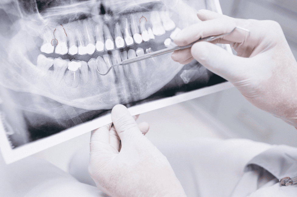
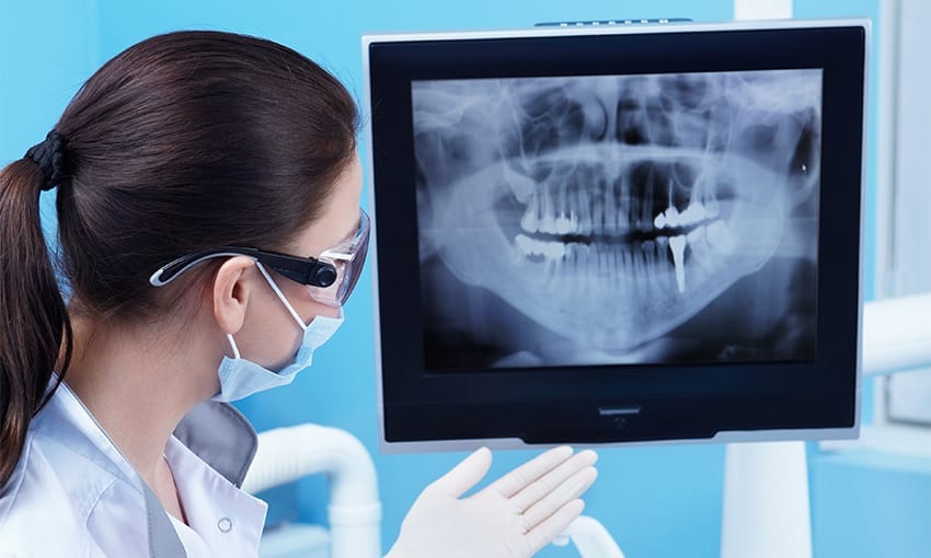
Radiographs have been a diagnostic tool for ages due to their accuracy and precision in identifying pathologies. Science and technology have evolved tremendously over the recent decades and invented several innovative advancements, replacing traditional computed tomography with cone beam computed tomography (CBCT)
The dentist in Buffalo Grove offers state-of-the-art cone beam CT which produces 3D images for accuracy. Let’s explore cone beam CT further.
What is CBCT?
Cone beam computed tomography (CBCT) is a type of X-ray machine used in situations where a traditional dental X-ray is not sufficient. With a CBCT, a cone-shaped X-ray beam rotates around the patient’s head to produce between 150 to 200 high-resolution 2D images. These are then digitally combined to form a 3D image. With a CBCT, your dentist can review 3D cross-sections of your head and neck.
A CBCT can identify the diseases of the jaw, dentition, and bony structures of the face, nasal cavity, and sinuses much more efficiently.
What are the common uses of CBCT?
Dentists commonly use a CBCT for treatment planning of orthodontics and dental implants. However, CBCT is also useful for more complex cases like:
- Surgical planning of impacted teeth
- Diagnosing TMJ disorder
- Evaluation of the sinuses, jaw, nerves canals, and nasal cavity
- Detecting, measuring, and treating jaw tumors
- Determining bone structure and tooth orientation
- Locating the origin of the pain or pathology
- Cephalometric analysis
- Reconstructive surgery
- Accurate and safe placement of dental implants

How is the procedure performed?
You will be taken to a radiology room and asked to sit or stand. The dental assistant will position you in such a way that the area of concern is positioned in the center of the beam. You will be asked to remain still while the X-ray source and detector revolve around you for a 360-degree rotation or less.
The procedure typically takes around 15 to 20 seconds for a complete volume, known as a full-mouth X-ray. For a regional X-ray, it may take around 10 seconds.
What are the benefits of CBCT?
CBCT is preferred over traditional CT due to the following benefits:
- It is fast and non-invasive
- Provides 3D images
- The radiation dosage is low
- The images are more accurate
Conclusion
Early and prompt detection of dental pathologies paves the way for precise and effective treatment plans. This can decide the fate of treatment outcomes. CBCT is a modern way of dental imaging that has effectively replaced traditional CT which helps in a variety of cases like orthodontic evaluation and implant placement.


